 Maureen Kohi, MD |
UCSF’s study sought to evaluate whether targeting only the vascular pedicles of the fibroid would significantly decrease the fibroid volume, thereby reducing procedural time. The idea is based on the principles employed in uterine fibroid embolization, another treatment method.
Patient recruitment turned out to be very challenging, even in a large population center like San Francisco. Although the study design called for six patients, only four were selected for this protocol. It was difficult to identify patients with the appropriate fibroid characteristics who were willing to undergo repeated imaging prior to treatment.
The procedural time for all patients was one hour, which is considerably less than the three to four hours required for the approved focused ultrasound procedure. All patients had excellent fibroid de-vascularization, with a non-perfused volume (NPV) to total fibroid volume ratios of 73%, 68%, 65%, and 55%.
Three months after the procedure, all patients experienced a reduction in fibroid-related symptoms and an improvement in quality of life. Three of the four patients opted for repeat focused ultrasound to completely ablate the remaining fibroid tissue. After one year, the remaining patient underwent myomectomy for persistent symptoms.
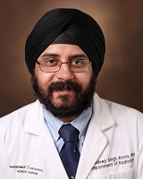
 Sandeep Arora, MBBS |
Vascular targeting during focused ultrasound may result in shorter procedural time, greater fibroid de-vascularization, and symptom improvement, but further research should quantify the amount of de-vascularization necessary to create high NPVs, alleviate symptoms, and prevent the need for additional treatments.
Dr. Arora is now a body imaging and interventional radiologist at Vanderbilt University Medical Center, and has joined the focused ultrasound research group there. “I would like to thank the Foundation and Dr. Kohi for their support, as this was the first research project in therapeutic ultrasound that I was involved in and has helped me to launch my research career,” he said.
