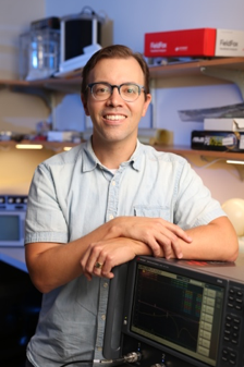
 Will Grissom, PhD, Associate Professor of Biomedical Engineering at Vanderbilt University, recently completed a year-long, remote-based research fellowship with the Foundation. He researched and developed new methods for measuring brain temperature during focused ultrasound treatments, resulting in the publication of two manuscripts and the completion of a collaborative multisite project. Read below to learn about the three projects wherein Dr. Grissom collaborated with Foundation scientists and focused ultrasound experts throughout the greater community to improve imaging used to guide focused ultrasound.
“My fellowship projects have the potential to greatly impact the field,” said Grissom. “We continue to work to develop imaging tools and improvements that improve patient care and enable focused ultrasound to treat more regions of the brain.”
“This fellowship is an example of the kind of collaboration the Foundation is catalyzing to accelerate development and adoption of focused ultrasound,” said John Snell, PhD, the Foundation’s Brain Program Technical Director. “Although Will made several trips to Charlottesville to work in the UVA Focused Ultrasound Center, he worked from Vanderbilt most of the time.”
The sections below describe the three projects led by Dr. Grissom during his Fellowship year.
Project 1: Improve the quality of MRI brain images used by physicians during transcranial focused ultrasound treatments.
Temperature monitoring is important when using focused ultrasound for thermal ablation, and the ability to accurately measure temperature is a critical part of the treatment process. Currently, MRI temperature imaging captures one thin slice of the brain every three seconds. The goal of this project was to expand the volume of tissue temperature mapping without sacrificing the rate of imaging to simultaneously image multiple slices without more encoding. “We developed a method to pre-calibrate the slices ahead of time, then fit those reference images to the data measured during therapy and extract separate temperature maps for each slice,” said Grissom. “This mathematical fitting procedure creates temperature maps across three slices in the same time it used to take to get one.”
Expanding the temperature imaging volume improves safety by allowing the treating physicians to visualize temperatures at the treatment site and in the rest of the brain. Simultaneous treatment monitoring and safety monitoring would improve patient care not only for the brain, but in other systems too. “We are going to adapt the method for treating prostate tissue, in collaboration with Profound Medical,” said Grissom. “Improving temperature monitoring allows faster and more precise treatments.”
See “Simultaneous Multislice MRI Thermometry with a Single Coil Using Incoherent Blipped‐controlled Aliasing” in Magnetic Resonance in Medicine >
Project 2: Reduce MRI image degradation caused by the presence of the water bath that is used to cool the skull during focused ultrasound treatments.
Treating the brain with focused ultrasound currently requires the use of a circulating water bath to effectively transmit the ultrasound to the brain and to prevent scalp heating. When the Insightec system begins a diagnostic MRI scan for anatomic or thermal imaging, it shuts off the circulation–but the water is still turbulent and keeps flowing for a while. This water movement can impact MRI images because it creates unpredictable bright and dark artifacts in the images of the brain, and the artifacts can then potentially degrade all subsequent temperature measurements.
To solve this challenge, Grissom worked with a postdoctoral colleague at the University of Virginia (Steven Allen, PhD) to modify the program that the MRI scanner runs to create temperature maps. Their idea was to select a limited part of the slice so that only the brain signals, and not the water bath signals, would appear in the final image. The duo developed a special radiofrequency pulse to suppress the water bath signal, creating a simple solution that significantly improves image quality while reducing temperature errors.
See “Reducing Temperature Errors in Transcranial MR-guided Focused Ultrasound Using a Reduced-field-of-view Sequence” in Magnetic Resonance in Medicine >
Project 3: Test a new MRI scanning approach to increase the volume of brain tissue that can be monitored for temperature during focused ultrasound.
As mentioned in Project 1, MRI temperature monitoring over the entire brain is a current challenge. After a 2016 brain workshop, the Foundation challenged several research groups to develop and propose new MR scan techniques to meet this need. Dr. Grissom’s final project was to compare the submitted sequences to determine which method best met the technical and clinical requirements established at the workshop.
Read more about this Volumetric Thermometry Challenge >
Related Stories
Vanderbilt’s Will Grissom Joins Foundation Fellowship Program September 2018
Foundation Funded Research Update – MR Thermometry October 2016
Open Source Focused Ultrasound System Now Available to Researchers Around the World May 2016
Q & A with Vanderbilt University’s Will Grissom and Charles Caskey May 2016
Vanderbilt’s Grissom Receives External Research Award to Improve Brain Temperature Imaging April 2014
