Key Points
- Christoph A. Binkert, MD, MBA, of Kantonsspital Winterthur in Switzerland, shares a report of a patient with sacroiliac joint (SIJ) osteoarthritis.
- Learn how focused ultrasound therapy helped the patient control her disabling pain and return to work.
The Foundation thanks Christoph A. Binkert, MD, MBA, Director of the Institute for Radiology and Nuclear Medicine and Head of the Clinic for Interventional Radiology and Vascular Surgery at Kantonsspital Winterthur in Switzerland, for providing this case report.
The Patient
 A 53-year-old female with severe, bilateral lower back pain presented to the clinic. The pain occasionally radiated into her legs and was more severe on the right side. It was persistent over several years but worsened over the past year. The pain was exacerbated by standing for long periods of time, which was especially cumbersome, given her career as a bakery owner.
The patient rated the low back pain a 70 out of 100 on a visual analog scale. To control the pain, she took a combination of medications, including non-steroidal anti-inflammatory drugs (NSAIDs) multiple times per day and Tramadol once per day.
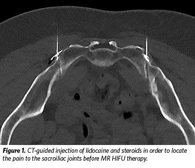
On clinical exam, there was the suspicion of sacroiliac joint (SIJ) osteoarthritis. To confirm the diagnosis, CT-guided SIJ injections (Fig 1) were performed bilaterally using a steroid combined with lidocaine (40 mg triamcinolone and 2 ml of lidocaine 2% on each side).
After the injection, the pain was markedly improved for two weeks with a slow relapse of pain thereafter. The patient was then sent to the interventional radiology (IR) clinic for MR-guided high intensity focused ultrasound (MR-HIFU) evaluation.
The Procedure
For a safe MR-HIFU intervention, it is important that the patient remains completely still during the sonications; therefore, spinal anesthesia or general anesthesia is necessary. In this case, the patient preferred general anesthesia, which was accomplished with a laryngeal mask approach.
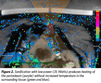 The patient was then positioned in a supine position on the MR-HIFU table. Sonications were performed with the following parameters: 4 mm-sized treatment cells, a transducer frequency of 1.2 MHz, and a power of 30 to 70 Watts. In total, seven sonications around the left SIJ and six around the right SIJ were completed. Temperature rise was observed during the thermal ablation with MR imaging (Fig 2).
The patient was then observed in the clinic and discharged home after two hours of observation.
Follow-up
At her six-week follow-up visit in the IR clinic, the patient reported that her low back pain had completely subsided on the left side within two days of the procedure and there was mild residual pain on the right side. Her pain level decreased to 21 out of 100 on a visual analog scale. The amount of pain medication she required was reduced to one dose of NSAIDs per day and Tramadol only occasionally, about once per week.
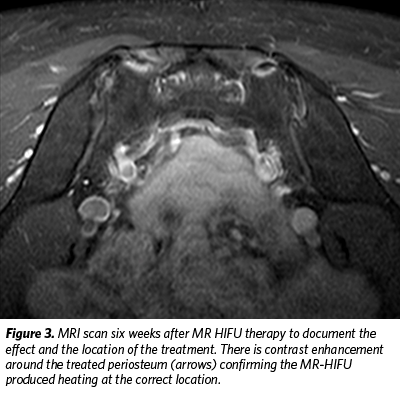
An MRI performed at the six-week follow-up revealed typical findings after MR-HIFU treatments of SIJs: small areas of contrast enhancement around the periosteum at the treatment site (Fig 3).
 The patient recently came to her six-month follow-up appointment with no pain on either side, and she takes no more pain medication. The patient was happy with the result, especially because working as a baker is now possible again.
Discussion
Chronic lower back pain is a common disease in the Western world. Osteoarthritis of the SIJ is responsible for about 15% to 30% of cases. There are often no specific radiological findings for SIJ osteoarthritis; therefore, CT-guided injections to test for pain relief are standard practice to confirm the diagnosis before HIFU treatment. The correct diagnosis prior to MR-HIFU therapy is important for success.
If conservative therapy fails, treatment options for SIJ osteoarthritis are limited. Arthrodesis is an invasive surgery, and minimally invasive radiofrequency ablation (RFA) carries risk associated with breaking the skin barrier and can be rather expensive due to the cost of the RFA probes. Therefore, MR-HIFU is a potentially attractive therapeutic option that is entirely noninvasive. In our experience, the procedure can be performed on an outpatient basis, and the results are promising.
Reference
Najafi A., Sartoretti E., Binkert CA. Sacroiliac Joint Ablation Using MR-HIFU. Cardiovasc Intervent Radiol (2019) 42:1363–1365