 An international group of researchers has designed, implanted, and completed preclinical testing of a biocompatible cranial prosthesis for improving imaging and therapeutic ultrasound transmission across the skull. Their work, “In Vitro and In Vivo Characterization of a Cranial Window Prosthesis for Diagnostic and Therapeutic Cerebral Ultrasound,” was recently published in the Journal of Neurosurgery.
The research was led by Francesco Prada, MD, a neurosurgeon at Istituto Neurologico Carlo Besta in Milan, Italy, and Wynn Legon, PhD, Assistant Professor in the Department of Neurological Surgery at the University of Virginia (UVA). Other Foundation collaborators included Brain Program Project Engineer, David Moore, and Director of Applied Physics Research, Frederic Padilla, PhD.
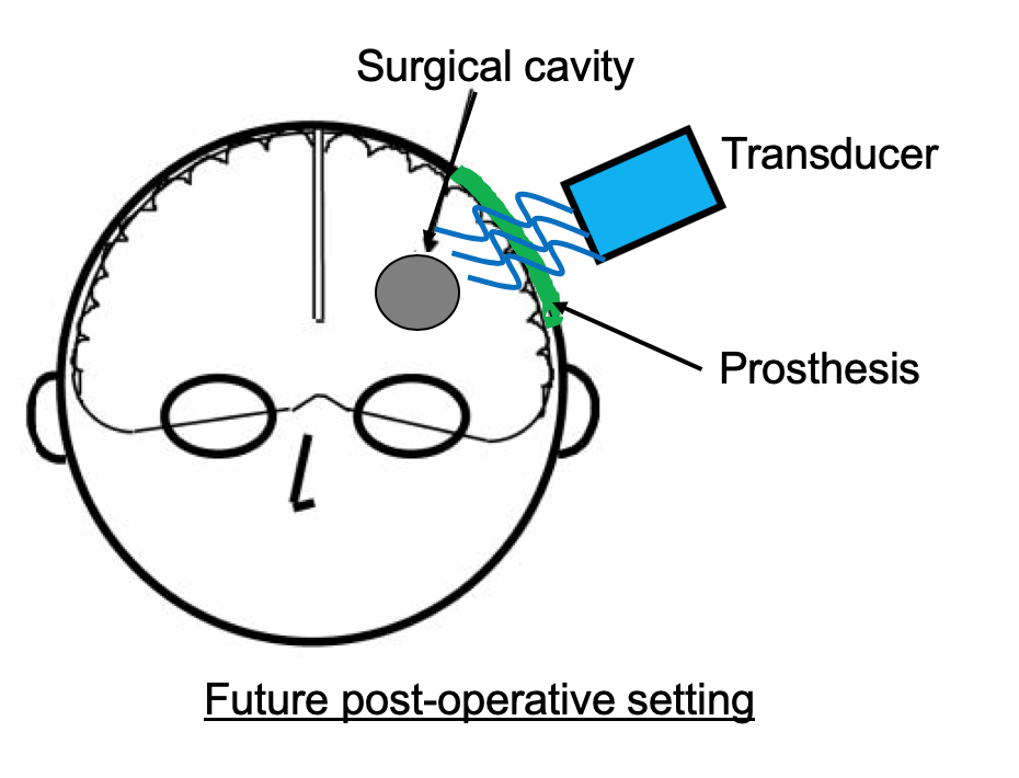
The prosthesis was designed to improve the diagnostic and therapeutic capabilities of ultrasound by decreasing its attenuation as it crosses the skull. It is a dense, low-porosity polyolefin-based polymer with favorable acoustic properties. Its homogeneous artificial structure creates consistent and predictable interactions with acoustic energy beams.
The testing processes confirmed that the novel prosthesis created an effective window for delivering focused and unfocused ultrasound and for obtaining intracranial ultrasound images.
 This technique was patented by Prada’s site in Milan, but the research was conducted at the UVA during the two years Prada spent as the Foundation’s first clinical Merkin scholar and then as Brain Program Director. During this time, he also served as Assistant Professor at UVA, where he collaborated with Legon.
“As a neurosurgeon, every time we perform a craniotomy, we carve a window for ultrasound,” explains Prada. “Since many craniotomies are performed every day around the world – a practice that will continue for the foreseeable future – we should take advantage of this opportunity. Implanting an ultrasound-transparent prosthesis at the end of the procedure will allow us to diagnose and treat the brain using ultrasound, even in the postoperative period, as is already the standard for many other organs.”
Building upon this data, the team plans to do in-vivo studies to further understand the acoustic properties of the prosthesis, perform ultrasound-guided focused ultrasound treatments, and ultimately bring the device in a clinical setting.