Key Points
- A multisite clinical trial is investigating focused ultrasound for patients with Alzheimer’s disease.
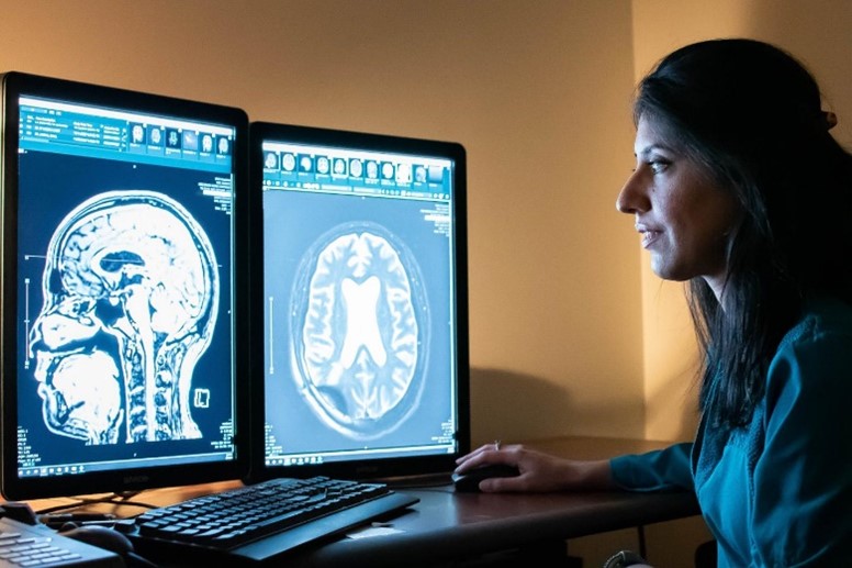
- Radiologists at West Virginia University evaluated contrast agent enhancement patterns from three trial participants during and after treatment.
- Post-treatment images revealed a unique contrast enhancement pattern downstream from targeted sites.
Blood-Brain Barrier Opening with MRI-guided Focused Ultrasound Elicits Meningeal Venous Permeability in Humans with Early Alzheimer Disease
 Researchers at West Virginia University’s Rockefeller Neuroscience Institute are participating in a multi-site clinical trial using focused ultrasound to temporarily and reversibly open the blood-brain barrier in a part of the brain called the hippocampus in patients with Alzheimer’s disease.
Radiologists at WVU sought to evaluate the contrast agent enhancement patterns from three trial participants during and after the treatment. They found post-treatment images revealed a unique contrast enhancement pattern downstream from targeted sites.
“The glymphatic system, which is a fluid-movement and waste-clearance system that’s unique to the brain, has been studied in animals, but there is controversy about whether this system truly exists in humans,” said lead author, Rashi Mehta, MD. “The imaging pattern that we discuss in the paper offers evidence not only to support that the system does likely exist in humans, but that focused ultrasound may modulate fluid movement patterns and immunological responses along this system.”