After reading his list of current projects, it is easy to see why Eli Vlaisavljevich, PhD, an Assistant Professor in the Department of Engineering and Mechanics at the Virginia Tech – Wake Forest School of Biomedical Engineering and Sciences, jokes that his Therapeutic Ultrasound and Non-Invasive Therapies Laboratory should perhaps be called “the unfocused focused ultrasound lab.” With a groundbreaking clinical trial named in memory of his mother and work ranging from cancer to conservation, Dr. Vlaisavljevich shows no signs of slowing down or focusing on only one idea anytime soon. Our interesting Q&A session with him follows.
Focused Ultrasound Work
When and how did you become interested in focused ultrasound?
I was an undergraduate biomedical engineering student at Michigan Tech doing work in biomaterials.* I had just been awarded a National Science Foundation graduate scholarship with the intention of pursuing graduate research in a biomaterials lab when my undergraduate advisor, Dr. Rupak Rajachar, invited a guest speaker to our class who was his good friend from the University of Michigan (UM). Zhen Xu, PhD, presented some of the first work being done with histotripsy, and I can still visualize her slide — the exact video she showed — of histotripsy boring a hole in cardiac tissue. I always wanted to work in a field that affected patient outcomes and quality of life, and the idea of noninvasive tissue removal really caught my attention. Like most people, I have lost family members to cancer, so I understood the idea of using histotripsy to treat cancer right away. I remember telling Zhen, “I have no background in acoustics, but I’m really interested in your work.” I think my advisor had to convince her to take me on as a graduate student, but I am thankful that she gave me a chance, because it was an incredible experience to go to graduate school at UM and work in the histotripsy group laboratory. I completed both my master’s and doctoral degrees there, so all three of my degrees were in biomedical engineering.

How did you decide to take your current position at Virginia Tech (VT)?
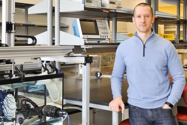
I completed my postdoctoral fellowship partly at UM and partly at HistoSonics, which was a great experience as we were working to advance histotripsy into a first clinical trial for treating liver cancer. Although I continue to enjoy my close partnership with HistoSonics, I have always felt more at home in the academic path where I can work with students and develop new technologies. Although I looked at several amazing opportunities, I accepted the position in the department of Biomedical Engineering and Mechanics (BEAM) at VT because it was the best fit for me. It has a great location close to the Focused Ultrasound Foundation and other major partners for my lab. The reputation of our engineering department, especially with ablation technologies, is superb–including the work that is being done here by Rafael Davalos, PhD, one of the leaders in irreversible electroporation (another type of nonthermal ablation that is also showing great results when trying to induce an immune response). VT also has great infrastructure, incredible partnerships, a growing focus on biomedical research, and an amazing interdisciplinary approach to research. I am able to do a lot of work with the veterinary school, and that was a big pull for me. We are not only developing animal models; we are also treating companion animals with our technologies, such as dogs and cats, that have diseases that are similar to humans. My lab is called the Therapeutic Ultrasound and Non-Invasive Therapies Laboratory.
Editor’s note: Dr. Vlaisavljevich discloses that he has a research partnership and financial relationship with HistoSonics.
What is the goal of your work?
The overarching goal of our laboratory is to investigate the physical mechanisms with which ultrasound interacts with different tissues in order to develop noninvasive therapies for a wide range of clinical applications.
What are your areas of interest in focused ultrasound?
As described on our website, our work is separated into five primary research areas:
- Histotripsy (Noninvasive Tissue Ablation)
– Cavitation physics
– Effects of tissue properties on histotripsy
– Device development for specific clinical applications - Acoustically Active Biomaterials
- Ultrasonic Neuromodulation
- Ultrasound-guided Tissue Regeneration
- Biomedical Technologies for Conservation
What mechanisms and clinical indications do you study?
This is the section about having an unfocused focused ultrasound lab – our projects are broad in scope and are targeting many clinical indications in oncology as well as other areas.
Because of my background with histotripsy, it is the primary mechanism that we study and what my lab is set up for. We are developing histotripsy for the treatment of multiple cancer types, including liver, kidney, pancreatic, soft tissue sarcoma, osteosarcoma, breast, and brain cancers. There is really no tumor that I’m not going to go after with histotripsy eventually if I get the chance. A large part of this work is developing tissue-specific treatment methods that can allow histotripsy to selectively ablate tumors in high-risk locations near critical structures, such as bile ducts, nerves, and blood vessels as well as ablate more fibrous tissues, such as cholangiocarcinoma liver tumors, which have recently been a particular focus for our group.
We are also working with Dr. Coy Allen on using histotripsy to induce immunomodulation. Our goal is to answer the key preclinical questions necessary to demonstrate the long-term oncological and immunological responses of different cancer types to histotripsy in order to advance this technology into a broader patient population and curative endpoints.
We have separate projects, including a grant to work with Adam Maxell and the University of Washington group, to develop novel “dual-modality” therapy methods that combine histotripsy with thermal ablation for the treatment of stiff tissues, such as cholangiocarcinoma, prostate, and uterine fibroids. We want to use controlled focused ultrasound to thermally modulate the tissues’ mechanical properties and then use histotripsy to effectively remove the fibrous tissue.

In veterinary medicine, our lab is using histotripsy to treat (both curative and palliative) naturally occurring cancerous tumors in dogs. Our collaboration with the Virginia-Maryland College of Veterinary Medicine is investigating this technology for osteosarcoma (PI: Dr. Joanne Tuohy) and soft-tissue sarcoma (PI: Dr. Shawna Klahn). We are also working closely with the histotripsy group at UM and Dr. Francesco Prada on a brain tumor study in dogs. This project, which is led by Dr. John Rossmeisl, is investigating histotripsy for brain tumor ablation through an acoustically transparent cranial implant that was developed by Dr. Prada and his team. In addition to addressing high canine mortality associated with these types of tumors, we hope that this work will also translate to use in humans.
Nanoparticle-mediated histotripsy (NMH) is another major focus of my research program. Our current NMH projects are focused on developing methods for the selective ablation of breast and brain cancer metastases, which can be aggressive, multifocal, and diffuse. We are working closely on these projects with the Allen and Munson labs at VT as well as Dr. Yasemin Yuksel Durmaz at Istanbul Medipol University in Turkey. Yasemin has been my main partner on NMH since our time together at Michigan, and it is exciting to be continuing these studies together in our new locations. NMH uses acoustically active nanoparticles to decrease the cavitation threshold and create cavitation in selected regions of multifocal tumors and micro-metastases. We are also using the NMH ablation platform to investigate nanoparticle-guided strategies for targeted drug delivery and immunomodulation, and we recently started working with Dr. Sasha Klibanov at the University of Virginia (UVA) on microbubble-mediated histotripsy approaches.
For neuromodulation, we are collaborating with researchers on the UVA (Dr. Wynn Legon) and VT (Dr. Sarah Clinton) campuses to develop new transducers and treatment methods for various brain disorders, particularly depression. Using experimental platforms to study neuromodulation at the cellular and systemic levels, we are trying to understand the mechanisms by which ultrasound can activate or inhibit neural activity (e.g., novel pulsing patterns for reproducibly stimulating or blocking neural activity). We hope to determine the specific mechanisms underlying previously observed bio-effects and then develop improved methods for treating depression and other neurological disorders.
Our current biomaterials work started with a collaboration that began when the Focused Ultrasound Foundation funded a global intern, Kaylee Meyers, to work in our lab. Her project was to develop hydrogels that release nitric oxide on demand using focused ultrasound. Kaylee is an undergraduate student at Michigan Tech, but during her funded summer internship in my lab, she was able to collect the data here at VT. That initial work led to the receipt of an NIH R15 award, which is allowing her to continue the focused ultrasound studies at Michigan Tech. In addition to this project and the NMH studies above, we are also pursuing the development of some other acoustic biomaterials for a range of applications, including an ongoing study to develop an improved water coupling bath for MR-guided focused ultrasound treatments, which is currently being led by Dr. Steven Allen and conducted in collaboration with the Meyer lab at UVA and the Davis lab at VT.
For conservation, we were recently fortunate to get a grant from the Gordon and Betty Moore Foundation to develop ultrasonic methods for enhancing DNA extraction, improving infectious disease screening, preventing the illegal trafficking of protected plants and animals, and tracing supply chains through species-specific identification methods. Our part of this collaborative project between our lab and Conservation X Laboratories is to improve DNA extraction methods by using acoustic cavitation clouds to break up complex cellular structures and release more DNA. Many of the samples we are focused on, such as timber, have only small amounts of DNA that is difficult to extract, and these tests often need to be done in remote locations away from the laboratory. If successful, this work has the potential to protect endangered species, reduce poaching, and improve diagnostic and therapeutic tools that can hopefully be used in medical field as well.
What are your funding sources?
- VT Institute for Critical Technology and Applied Science Carilion Clinic
- Edward Via College of Osteopathic Medicine
- National Institutes of Health
- iThriv, the Integrated Translational Health Research Institute of Virginia
- Focused Ultrasound Foundation
- The Gordon and Betty Moore Foundation
- The American Kennel Club
- National Geographic
Research Details

Who are your team members?
I am very fortunate to be working with a large and diverse group of undergraduate and graduate students as well as many internal and external research collaborators. Right now, our main group includes eight graduate students: six doctoral and two masters. We have nine undergraduate engineering students. Promoting undergraduate-level research is important to me and to the School of Engineering. We also have eight medical students from the VT Carillon Medical School, including Pete Weber, who previously worked at the Focused Ultrasound Foundation. Pete has been working on our histotripsy cholangiocarcinoma project, which is funded by the Foundation. The medical students split time between Blacksburg and Roanoke. Hal Homes, PhD, is a part time research scientist in our lab in addition to his role as lead engineer at Conservation X Labs located in Seattle, Washington. I am also currently advertising for a postdoctoral fellow and a few other positions so hopefully you can spread the word if anyone is interested.

Who are your internal and external collaborators?
We are a truly interdisciplinary lab, so we are fortunate to have a multitude of great partners. Being a product of the focused ultrasound community also creates many collaborations.
Internal at VT
- Irving Coy Allen, MS, MBA, PhD, Department of Biomedical Sciences & Pathobiology
- Joanne Tuohy, DVM, PhD, Department of Small Animal Clinical Sciences
- Shawna L. Klahn, DVM, Department of Small Animal Clinical Sciences
- Nick Dervis DVM, PhD, Department of Small Animal Clinical Sciences
- John Rossmeisl, Jr., DVM, MS, Department of Small Animal Clinical Sciences
- Shima Shahab, PhD, Department of Mechanical Engineering
- Jason Holliday, PhD, Department of Forestry Resources and Environmental Conservation
- David Luyimbazi, MD, Department of Surgery, Carilion Clinic
- Fidel Valea, MD, Department of Obstetrics and Gynecology, Carilion Clinic
- Jayasimha Rao, PhD, Department of Infectious Diseases, Carilion Clinic
- Jennifer Munson, PhD, Department of Biomedical Engineering and Mechanics
- Vincent Wang, PhD, Department of Biomedical Engineering and Mechanics
- Raffaella De Vita, PhD, Department of Biomedical Engineering and Mechanics
- Scott Verbridge, PhD, Department of Biomedical Engineering and Mechanics
- Rafael Davalos, PhD, Department of Biomedical Engineering and Mechanics
- Pamela VandeVord, PhD, Department of Biomedical Engineering and Mechanics
- John Robertson, Department of Biomedical Engineering and Mechanics
- Richey Davis, PhD, Department of Chemical Engineering
- Andre Muelenaer, MD, Department of Biomedical Engineering and Mechanics
- Abby Whittington, PhD, Department of Chemical Engineering
- Susan Campbell, PhD, School of Neuroscience
- Sarah Clinton, PhD, School of Neuroscience
External
- Zhen Xu, PhD, and Tim Hall, PhD, University of Michigan Department of Biomedical Engineering
- Fred Lee, Jr., MD, and Tim Ziemlewicz, MD, University of Wisconsin Department of Radiology
- Adam Maxwell, PhD, and George Schade, MD, University of Washington Department of Urology
- Joan Vidal-Jove, Khuab Institute for Interventional Oncology, Barcelona, Spain
- Rupak Rajachar, PhD, Michigan Tech Department of Biomedical Engineering
- Mishal Mendiratta-Lala, MD, University of Michigan Department of Radiology
- Wynn Legon, PhD, UVA
- Steven Allen, PhD, Brigham Young University
- Yasemin Yuksel Durmaz, PhD, Istanbul Medipol University
- Jeremy Brown, PhD, Dalhousie University Department of Biomedical Engineering
- Francesco Prada, MD, Carlo Besta Neurological Institute, Milan, Italy
- Sasha Klibanov, PhD, UVA
- Eric Johnsen, PhD, University of Michigan Department of Mechanical Engineering
- Ken Bader, PhD, University of Chicago
- Amanda Smolock, MD, PhD, Medical College of Wisconsin
- Wake Forest Comprehensive Cancer Center
- Children’s National Hospital
- HistoSonics
- Daxsonics
- Conservation X Labs
- Intelligenza Trasparente
For both internal and external, I listed the main people I could think of – but there are of course others that aren’t listed, especially in cases like Wisconsin, Michigan, and Washington where we are working with big teams.
What are your greatest achievements? Any major disappointments?
Working with HistoSonics and helping launch its first clinical trial for liver cancer was very personal to me, because the THERESA study was named after my mom, who died from liver cancer when I was 4 years old. I would say this is my biggest accomplishment to date because of the impact it will hopefully have on actual patients.
The biggest disappointment in my career so far was likely when I embarrassed myself in graduate school by trying to use histotripsy to scramble an egg inside the shell. I made a big deal about how it would work, but I was completely embarrassed in front of everyone when we couldn’t generate a bubble cloud inside the shell and an unscrambled egg came out. Our histotripsy technology has come a long way since then, so I am sure we will revisit that application one day, but for now it remains a big disappointment that I am still trying to live down.
What do you see as impediments to your success?
There are obviously many challenges that are unique to each application that we are working on. If I had to pick one overall impediment that comes up most often, I would have to say improving the imaging methods we use for guiding and monitoring our treatments. This is something we are working through in our pancreatic histotripsy studies in which our therapy has the ability to treat with high precision, but we are limited by the quality of our ultrasound imaging in visualizing the pancreas for targeting. Similar challenges are seen in other applications, which is why this remains a large focus area for my group and others.
What is on your research wish list?
I feel fortunate for my current role and the ability to be where I am with our great partnerships and collaborations, so I wouldn’t want to test my luck by wishing for too much more. That said, my main wish is for each student that works in our lab to see their efforts translate into some clinical impact either in the short or long term.
In the next 5 to 10 years, I would love to walk through a hospital that I’m not associated with and see a histotripsy system sitting outside one of the rooms. That’s a moment that I’m excited for. I’ve seen one of our systems in a clinic for a clinical trial, but I haven’t yet seen a commercial system in use somewhere. It would be cool to run into one randomly. This is also true for veterinary histotripsy systems that I’d like to see deployed across the globe.
I also would love to hear a person say that someone in their family received some type of “histo” treatment — they don’t even need to remember the name of it — and it had a great effect on that person. In the veterinary realm, I am looking forward to seeing previously sick and limping dogs with osteosarcoma running around as happy, pain-free dogs after undergoing a successful histotripsy procedure without the need to take their limbs.
Finally, from a student perspective, I am looking forward to seeing our students graduate and move into academia, industry, and other opportunities as they continue to expand this amazing network and focused ultrasound community that we have all built together. I look forward to continuing to playing a small role in developing our community.
Has the Foundation played a role in your work?
The Foundation has played a major role in helping me establish my research program at VT. I have been extremely fortunate to have been awarded multiple research grants from the Foundation, including funding to study histotripsy for the treatment of cholangiocarcinoma tumors as well as a grant to establish a pig tumor model for studying histotripsy for the treatment of pancreatic cancer. In addition, we have recently received several grants for our veterinary clinical trials treating companion animals with soft tissue sarcoma, osteosarcoma, and brain tumors. However, beyond the direct research funding, the Foundation has played an even bigger role in helping me to establish my new lab by connecting me with other researchers, clinical collaborators, and industry partners. These connections have led to multiple exciting new projects in my lab that we wouldn’t be able to pursue without the support and connections of the Foundation.
Clinical Details
How many patients have you treated?
Although VT does not currently have any human clinical trials for focused ultrasound, HistoSonics enrolled eight patients in the THERESA study, which I was a part of both through my position at the company and later as a research collaborator. I am excited to see this work continue with their currently planned clinical trials that will extend histotripsy to additional patients.
In our current veterinary clinical trials, we have enrolled the first two companion animal patients for our soft tissue sarcoma and osteosarcoma trials. These studies are funded by the Focused Ultrasound Foundation and the American Kennel Club. We are in the process of planning for our first canine brain tumor treatments in early 2021. There was also a previous study here at VT using the Theraclion system for treating soft tissue tumors in 20 dogs, which was conducted by my veterinary colleagues in partnership with Theraclion and the Foundation.
Our medical students, in particular, are making the connection between laboratory research and clinical needs. Furthermore, our clinical collaborators in radiology, urology, and oncology truly help us identify the problems that need to be solved for bringing this technology into patient care.
Do you have any interesting research stories or highlights?
One interesting story from last year was a project we were close to starting with a wildlife ecologist in Tasmania who is studying Tasmanian devil facial tumor disease. It’s a contagious tumor that spreads when these animals bite each other, and it has decimated the Tasmanian devil population. I have always said that histotripsy is a platform technology with many different applications within the oncology field, but the thought of developing a treatment for Tasmanian devils was definitely not expected. We ended up having a couple of great conversations, and we were close to starting a research collaboration to do some feasibility studies when the COVID pandemic unfortunately put the project on hold. Nonetheless, I remain hopeful that we can revisit this in the future and add it to the growing list of indications for histotripsy! Until then, we have plenty to keep us busy.
*Dr. Vlaisavljevich also played college hockey at Michigan Tech!
Related Videos
Vlaisavljevich Research Laboratory | August 2019
Hal Holmes – Down to the Core – New DNA Technologies to Preserve Nature and Prevent Extinction | December 2019
Related Stories
Researcher Receives NIH Grant to Study Noninvasive Treatment for Metastatic Breast Tumors August 2020
3rd Quarter Research Awards September 2018